


Description
Specifications
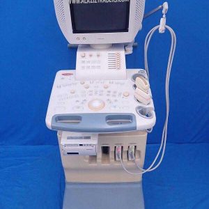
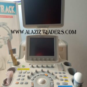
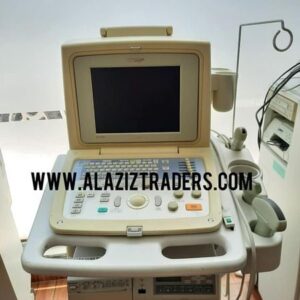
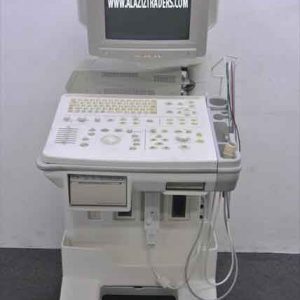
The GE Logiq S8 is an ultrasound that supports a variety of applications. Including cardiac, abdominal, obstetrics, gynecology, breast, small parts, urological, musculoskeletal, vascular, pediatric, neonatal, and transcranial. The system is compatible with over 27 different transducer/probes that deliver high-definition images. The Logiq S8 offers B-flow imaging, this can show blood flow visualization, where the system displays a grayscale or color images of the blood flow echoes. This system can help diagnose vessel wall irregularities, Carotid Plaque, Thyroid nodule activity, Kidney Perfusion, Vascular disease, and many others.
The GE Logiq S8 ultrasound system has a 23 inch LCD monitor and a 9 in touch screen for adjusting the imaging parameters. The lightweight, compact ultrasound system is easy to maneuver and move around to fit your ultrasound needs. The Logo S8 offers streamlined arc hinging and reporting, making it easy to connect to the facility’s computer system to view previous ultrasounds or transfer current ultrasound imaging and patient data.
- Advanced user interface with high resolution wide 9” LCD touch panel
- 4 Active probe ports and 1 parking
- Brightness & contrast adjustment
- Integrated gel warmer
- Power Assistant
- Maximum Depth of Field: 0 – 33 cm
- Virtual Convex
- SRI-HD (Speckle Reduction Imaging)
- CrossXBeam
- User Programmable Preset Capability
- Automatic Optimization
- Real-time Doppler Auto measurements/calculations
- Compare Assistant
- Raw Data Processing (All Imaging control parameters can be changed)
Dimensions
- Height: minimum 45.3 in (1150 mm)
- Height: Maximum 67.7 in (1720 mm)
- Width: casters 24.4 in (620 mm)
- Width: keyboard 19.7 in (500 mm)
- Depth: maximum 34.6 in (880 mm)
- Depth: casters 31.1 in (790 mm)
- Weight: 187.4 lb (85kg)
XDClear
- Digital Beamformer
- Displayed Image Depth: 0-33 cm
- Minimum Depth of Field: 0-2 cm (zoom) probe dependent)
- Maximum Depth of Field: 0-33 cm (probe dependent)
- Contrnuous dynamic receive focus/continuous dynamic receive aperture
Electrical Power
- Voltage: 100-120 Vac or 220-240 Vac
- Frequency: 50.60 HzPower: Consumption maximum of 900 VA with peripherals
Image Storage
- On-board database of patient information from past exams
- Storage Formats
- DICOM – Compressed/ uncompressed, single / multifram, with/ without Raw Data
- Export: JPEG, JPEG2000, WMV (MPEG 4), and AVI formats
Storage Devices
- USB Flash Device: 64MB to 64GB (for exporting individual images/clips)
- CD-R storage: 700MB
- DVD storage: R (4.7GB)
- Hard Drive Image Storage: ~300GB
- Compare previous exam images with current exam images
- Reload of archived data sets
- Network Storage support for Import, Export, DICOM Read, SaveAs,
Reviews
There are no reviews yet.